


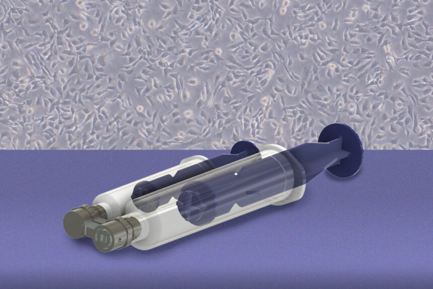
Researchers at the Massachusetts Institute of Technology and Johns Hopkins University developed a handheld device in September 2026 designed for early cancer detection by gently collecting living cells from specific tissue locations. Published in the journal Device, the technology aims to help doctors detect microscopic precursor lesions of high-grade serous ovarian cancer much earlier, potentially improving patient survival rates when caught before advanced stages.
How does the cell-collection device work?
The handheld system utilizes a 3D-printed microfluidic channel and a vacuum seal to apply controlled fluid shear stress to specific tissue regions. This process safely detaches viable living cells without destroying surrounding tissue or requiring harsh chemical preservatives, allowing scientists to cultivate the recovered cells into organoids for disease modeling and treatment testing.
Why does this medical advance matter?
Ovarian cancer is often diagnosed in later stages when survival rates drop significantly, largely because early microscopic precursor lesions in the fallopian tubes are difficult to sample. By enabling targeted cellular collection while preserving the underlying tissue for standard pathology, the breakthrough supports personalized medicine and earlier diagnostic capabilities for patients.